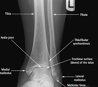
ANKLE ANTERO-POSTERIOR OR MORTISE PROJECTION
Ankle के दो रूटीन प्रोजेक्शन लिए जाते है anterio-posterior (AP, mortise view) तथा lateral projection | Mortise view, tibia तथा fibula के मध्य syndesmosis देखने के लिए किया जाता है |
Ankle में ankle injuries, including sprains and fractures के साथ साथ Syndesmosis ligament injuries भी पायी जाती है |
The syndesmosis is the name of the ligament that connects two bones of the leg. These bones, the tibia, and fibula are between the knee and ankle joints. The tibia is the larger shin bone that supports most of the weight of the body, and the fibula is the smaller bone on the outside of the leg. Connecting these bones is a ligament called the syndesmosis, also called the syndesmotic ligament.
Ankle में ankle injuries, including sprains and fractures के साथ साथ Syndesmosis ligament injuries भी पायी जाती है |
Position of patient and image receptor
पेशेंट को एक्सरे टेबल पर बैठाते है तथा leg को एक्सटेंड करते है |
Affected ankle को dorsiflexion के लिए एक firm 90° pad के द्वारा उसकी planter surface पर लगाकर सपोर्ट दिया जाता है |
Limb को इस स्थिति से लगभग 20° तब तक medially rotate किया जाता है जब तक medial तथा lateral malleoli इमेज रिसेप्टर से समान दुरी पर ना हो जाये |
अगर पेशेंट फुट को dorsiflex करने में अक्षम हो तो heel को 15° wadge की सहायता से ऊपर उठा देते है या एक्सरे बीम को 5°-10° cranial angle देते है |


कोई टिप्पणी नहीं:
एक टिप्पणी भेजें